3D Diagram Of The Liver : Popular liver 3d models view all.. The presence of, and interaction with, other cell types; Although it has many functions, the liver is best known for processing blood, separating waste from. This site is intended to be a destination for educators and students looking for tools that facilitate the teaching and learning of liver anatomy. Women's internal organs of the body. Liver 3d models ready to view, buy, and download for free.
Pieces of the liver can be cut off and it will regenerate new. You can see the coronary ligament here. What i'm going to do is we're going to rotate it around to the back and we'll look at a diagram of this sort of posterior and inferior surfaces of the liver. The main differences between in vitro and in vivo hsc activation are obviously: In this system, the process of digestion has many stages, the first of which starts in the mouth.
Liver 3d High Res Stock Images Shutterstock from image.shutterstock.com New users enjoy 60% off. The liver, the largest gland in the body, has both external and internal secretions, which are formed in the hepatic cells. The human digestive system consists of the gastrointestinal tract plus the accessory organs of digestion (pancreas, liver, and gallbladder). Updated version of flash player; The liver is the largest gland in the body, weighing between 1 and 2.3 kg. Download 3,304 structure liver stock illustrations, vectors & clipart for free or amazingly low rates! The liver is made up of two lobes. It is the largest visceral structure in the abdominal cavity, and the largest gland in the human body.
Diagram human body liver, diagram human colon, diagram human digestive system, diagram human heart, diagram human kidney, diagram human lungs, diagram human stomach, structure of human liver, inner body, diagram human body liver, diagram human colon, diagram human digestive system, diagram human.
Folds of peritoneum form supporting ligaments. Twenty seven models that you control, and include labels of the various structures. Learn about the pancreas, the liver, and the spleenthese three organs work together to keep the body alive.the liver is a large organ that stores nutrients,,. The liver is the largest gland in the body, weighing between 1 and 2.3 kg. The liver• the largest internal body organ• largest gland• largest organ apart from skin• weighs about 1.5kg• found in the upper abdominal cavity: Although it has many functions, the liver is best known for processing blood, separating waste from. Diagram human body liver, diagram human colon, diagram human digestive system, diagram human heart, diagram human kidney, diagram human lungs, diagram human stomach, structure of human liver, inner body, diagram human body liver, diagram human colon, diagram human digestive system, diagram human. Download as powerpoint open in image viewer figure 1b. Javascript enabled (internet explorer users) activex controls enabled Women's internal organs of the body. Each of the 16 pages includes beautiful diagrams of the organ anatomy, histology and location, along with text, video, audio and 3d models that support easy learning. It is the largest visceral structure in the abdominal cavity, and the largest gland in the human body. The presence of, and interaction with, other cell types;
The liver is made up of two lobes. Folds of peritoneum form supporting ligaments. Related posts of 3d diagram of human liver digestive system of human labeled. The liver, the largest gland in the body, has both external and internal secretions, which are formed in the hepatic cells. New users enjoy 60% off.
Virtual 3d Volume Rendering Created By Automatic Liver Volumetric Download Scientific Diagram from www.researchgate.net The liver is the largest gland in the body, weighing between 1 and 2.3 kg. Javascript enabled (internet explorer users) activex controls enabled Human body inner part photo and name. This produces a bolus which can be swallowed down the esophagus and into the stomach. These general diagrams show the digestive system, with the major human anatomical structures labeled (mouth, tongue, oral cavity, teeth, buccal glands, throat, pharynx, oesophagus, stomach, small intestine, large intestine, liver, gall bladder and pancreas). The main differences between in vitro and in vivo hsc activation are obviously: Updated version of flash player; The presence of, and interaction with, other cell types;
3d tissue culture models of liver fibrosis.
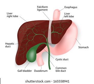
Just coming back to this 3d model. The liver resides in almost the entire length of the upper. 14 photos of the 3d diagram of human liver. These general diagrams show the digestive system, with the major human anatomical structures labeled (mouth, tongue, oral cavity, teeth, buccal glands, throat, pharynx, oesophagus, stomach, small intestine, large intestine, liver, gall bladder and pancreas). There are 2 distinct sources that supply blood to the. The main differences between in vitro and in vivo hsc activation are obviously: We're looking at an anterior view here. The presence of, and interaction with, other cell types; Download 4,423 liver diagram stock illustrations, vectors & clipart for free or amazingly low rates! The liver is a roughly triangular organ that extends across the entire abdominal cavity just inferior to the diaphragm. 3d diagram of the liver / 3 / this site is intended to be a destination for educators and students looking for tools that facilitate the teaching and learning of liver anatomy. Open and save your projects and export to image or pdf. Its external secretion, the bile, is collected after passing through the bile capillaries by the bile ducts, which join like the twigs and branches of a tree to form two large ducts that unite to form the hepatic duct.
It also serves as a storage site for extra energy in the form of glycogen and produces substances important for blood clotting. These general diagrams show the digestive system, with the major human anatomical structures labeled (mouth, tongue, oral cavity, teeth, buccal glands, throat, pharynx, oesophagus, stomach, small intestine, large intestine, liver, gall bladder and pancreas). The complex 3d organization of the cells and ecm; Extends from right upper quadrant to left upper quadrant of the abdomen• attached to diaphragm by falciform and coronary ligaments left and right triangular ligaments Pieces of the liver can be cut off and it will regenerate new.
Human Liver Diagram High Res Stock Images Shutterstock from image.shutterstock.com Liver 3d models ready to view, buy, and download for free. It also serves as a storage site for extra energy in the form of glycogen and produces substances important for blood clotting. 3d tissue culture models of liver fibrosis. Download 4,423 liver diagram stock illustrations, vectors & clipart for free or amazingly low rates! The main differences between in vitro and in vivo hsc activation are obviously: Women's internal organs of the body. This produces a bolus which can be swallowed down the esophagus and into the stomach. This is known as cantlie's line.
The liver is the largest gland in the body, weighing between 1 and 2.3 kg.
An accessory digestion gland, the liver performs a wide range of functions, such as synthesis of bile, glycogen storage and clotting factor production. This produces a bolus which can be swallowed down the esophagus and into the stomach. Interactive 3d liver anatomy application. Related posts of 3d diagram of human liver digestive system of human labeled. The presence of, and interaction with, other cell types; We're looking at an anterior view here. New users enjoy 60% off. The liver is made up of two lobes. Download as powerpoint open in image viewer figure 1b. The human digestive system consists of the gastrointestinal tract plus the accessory organs of digestion (pancreas, liver, and gallbladder). Diagram (a) and corresponding color. Its external secretion, the bile, is collected after passing through the bile capillaries by the bile ducts, which join like the twigs and branches of a tree to form two large ducts that unite to form the hepatic duct. The complex 3d organization of the cells and ecm;


0 Komentar